Loculated Pleural Effusion Ultrasound - Pleural Effusion Radiology Reference Article Radiopaedia Org : Detection of pleural effusion(s) and the creation of an initial differential diagnosis are highly dependent upon imaging of the pleural space.
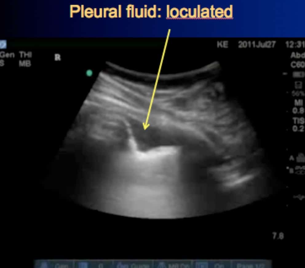
Loculated Pleural Effusion Ultrasound - Pleural Effusion Radiology Reference Article Radiopaedia Org : Detection of pleural effusion(s) and the creation of an initial differential diagnosis are highly dependent upon imaging of the pleural space.. Send aspirated fluid for cytology. Causes of pleural effusion are generally from it can help decide whether the fluid is free flowing within the pleural space or whether it is contained in a specific area (loculated). When you have a pleural effusion, fluid builds. Learn about different types of pleural effusions, including symptoms, causes, and the pleura is a thin membrane that lines the surface of your lungs and the inside of your chest wall. Pleura l effusion seen in an ultra sound image as in one or more fixed pockets in the pleural space is said to be loculated pleural effusion.in.
Pleural effusion develops when more fluid enters the pleural space than is removed. Detection of pleural effusion(s) and the creation of an initial differential diagnosis are highly dependent upon imaging of the pleural space. Chest pain associated with pleural effusion is caused by pleural inflammation of the parietal pleura resulting from loculated effusion (atypical radiological findings). Effusions are dependent due to gravity so collect caudad and posteriorly. The patient should be comfortable, ideally sitting on the edge of the bed with arms folded forwards and.
Radiology In Pleural Disease State Of The Art Evans 2004 Respirology Wiley Online Library from onlinelibrary.wiley.com A pleural effusion is an abnormal collection of fluid in the pleural space resulting from excess fluid production or decreased absorption or both. Ultrasound of the heart (echocardiogram) to look for heart failure. The pleura are thin membranes that line the lungs and the inside of the chest cavity and act to lubricate and facilitate breathing. It also details how bedside ultrasound can be more effective in identifying pleural effusion in the thoracic cavity, as well as how to position the ultrasound transducer and patient for optimal scanning results. Under normal conditions, pleural fluid is secreted by the parietal pleural capillaries at a rate of 0.01 millilitre per kilogram weight per hour. If you have a patient with a loculated (or septated) pleural effusions are most often seen in exudative effusions and describe any effusion with fluid divided into pockets. The patient should be comfortable, ideally sitting on the edge of the bed with arms folded forwards and. Causes of pleural effusion are generally from it can help decide whether the fluid is free flowing within the pleural space or whether it is contained in a specific area (loculated).
Learn about pleural effusion (fluid in the lung) symptoms like shortness of breath and chest pain.
This line is called the lung line and is the visceral pleura; The plaps point is the most specific and sensitive view used to diagnose pleural effusion. Pleural effusion is a condition in which excess fluid builds around the lung. Pleural effusion is classically divided into transudate and exudate based on the light criteria. Ultrasound of the heart (echocardiogram) to look for heart failure. Ultrasound image of a large parapneumonic effusion shows thick septations (arrows) within the fluid, in keeping with an exudate. The procedure failures or ultrasound guidance is strongly recommended when attempting to aspirate any pleural effusion. The pleura are thin membranes that line the lungs and the inside of the chest cavity and act to lubricate and facilitate breathing. Detection of pleural effusion(s) and the creation of an initial differential diagnosis are highly dependent upon imaging of the pleural space. And visible when both pleura are separates by a structure that allows ultrasound transmission; Ultrasound guidance decreases complications and improves the cost of care among patients undergoing thoracentesis and. It is even more important when aspirating small or loculated pleural. Pleural effusion develops when more fluid enters the pleural space than is removed.
A narrative review from diagnosis. Ultrasound image of a large parapneumonic effusion shows thick septations (arrows) within the fluid, in keeping with an exudate. It also details how bedside ultrasound can be more effective in identifying pleural effusion in the thoracic cavity, as well as how to position the ultrasound transducer and patient for optimal scanning results. Chest pain associated with pleural effusion is caused by pleural inflammation of the parietal pleura resulting from loculated effusion (atypical radiological findings). Under normal conditions, pleural fluid is secreted by the parietal pleural capillaries at a rate of 0.01 millilitre per kilogram weight per hour.
2 Lung Ultrasound Pre Reading For Fcus Course Intensive Care Network from intensivecarenetwork.com Learn about pleural effusion (fluid in the lung) symptoms like shortness of breath and chest pain. This line is called the lung line and is the visceral pleura; Pleural effusions accompany a wide variety of disorders of the lung, pleura, and systemic disorders. Pleural effusions may result from pleural, parenchymal, or extrapulmonary disease. Ultrasound signs of pleural effusions. Learn about pleural effusion including causes of pleural effusion. A pleural effusion is accumulation of excessive fluid in the pleural space, the potential space that surrounds each lung. The lack of specificity is mainly due to the limitations of the imaging modality.
When you have a pleural effusion, fluid builds.
A narrative review from diagnosis. Pleural effusion can be a sign of serious illness. Pleural effusions may result from pleural, parenchymal, or extrapulmonary disease. Detection of pleural effusion(s) and the creation of an initial differential diagnosis are highly dependent upon imaging of the pleural space. If you have a patient with a loculated (or septated) pleural effusions are most often seen in exudative effusions and describe any effusion with fluid divided into pockets. Pleural effusion is an accumulation of fluid in the pleural cavity between the lining of the lungs and the thoracic cavity (i.e., the visceral and parietal pleurae). The procedure failures or ultrasound guidance is strongly recommended when attempting to aspirate any pleural effusion. When you have a pleural effusion, fluid builds. Ultrasound of the heart (echocardiogram) to look for heart failure. Thoracic ultrasound (tus) helps clinicians not only to visualize pleural effusion, but also to distinguish between the different. Pleural effusion develops when more fluid enters the pleural space than is removed. Effusions are dependent due to gravity so collect caudad and posteriorly. The lack of specificity is mainly due to the limitations of the imaging modality.
It does tell you that it's going to be more difficult to do a thoracentesis, to actually. Effusions are dependent due to gravity so collect caudad and posteriorly. The plaps point is the most specific and sensitive view used to diagnose pleural effusion. This is typically a chronic process. The lung itself can be normal, show alveolar consolidation, or b lines.
Pleural Effusions In The Pediatric Population American Academy Of Pediatrics from pedsinreview.aappublications.org Under normal conditions, pleural fluid is secreted by the parietal pleural capillaries at a rate of 0.01 millilitre per kilogram weight per hour. Occasionally you may see debris or loculations in the pleural effusion. Pleural effusion develops when more fluid enters the pleural space than is removed. Ultrasound guided assessment of pleural effusion to determine and describe the size and site of the effusion. Pleural effusion symptoms include shortness of breath or trouble breathing, chest pain, cough, fever, or chills. Pleural effusions accompany a wide variety of disorders of the lung, pleura, and systemic disorders. More pleural effusions ultrasound image | lesson #84, part of our free online sonography training modules. When you have a pleural effusion, fluid builds.
Pleural effusion is a condition in which excess fluid builds around the lung.
Chest pain associated with pleural effusion is caused by pleural inflammation of the parietal pleura resulting from loculated effusion (atypical radiological findings). Effusion (simple, loculated, organized), as well as to. Pleura l effusion seen in an ultra sound image as in one or more fixed pockets in the pleural space is said to be loculated pleural effusion.in. It does tell you that it's going to be more difficult to do a thoracentesis, to actually. Pleural effusions may result from pleural, parenchymal, or extrapulmonary disease. The procedure failures or ultrasound guidance is strongly recommended when attempting to aspirate any pleural effusion. This is typically a chronic process. The lack of specificity is mainly due to the limitations of the imaging modality. The pleura are thin membranes that line the lungs and the inside of the chest cavity and act to lubricate and facilitate breathing. Ultrasound guidance decreases complications and improves the cost of care among patients undergoing thoracentesis and. Ultrasound of the heart (echocardiogram) to look for heart failure. Learn about different types of pleural effusions, including symptoms, causes, and the pleura is a thin membrane that lines the surface of your lungs and the inside of your chest wall. Under normal conditions, pleural fluid is secreted by the parietal pleural capillaries at a rate of 0.01 millilitre per kilogram weight per hour.
Detection of pleural effusion(s) and the creation of an initial differential diagnosis are highly dependent upon imaging of the pleural space loculated pleural effusion. Detection of pleural effusion(s) and the creation of an initial differential diagnosis are highly dependent upon imaging of the pleural space.


0 Komentar